spheroONE
Gentle, Image-Based Sorting and Isolation of 3D Cell Models for Smarter Drug Discovery
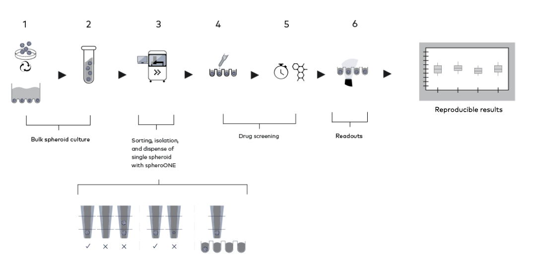
spheroONE is redefining how 3D cellular models are prepared and used in drug discovery. Its gentle sorting reduces manual handling and ensures every assay starts with homogenous spheroids or organoids. By transforming variability into consistency, spheroONE enhances data quality, accelerates timelines, and de-risks critical preclinical decisions.
spheroONE ensures consistent and functional 3D models by combining precise image-based isolation with gentle dispensing, based on user-defined parameters. It minimizes variability, preserves fragile structures, and adapts seamlessly across spheroids, organoids, and patient-derived models. With integrated brightfield and fluorescence imaging, it enables sorting based on morphology or phenotypic response. High speed and automation further reduce hands-on time, letting researchers generate reliable, reproducible data at scale with minimal manual intervention.
You're in good company
These leading institutions use our single cell and 3D cell model technologies:


Image-based 3D cell model sorting
Gentle isolation and dispensing

Selection by size morphology, or fluorescence

Controlled number of -oids per well
90% single 3D model accuracy
")
Automated and flexible
Benefits
spheroONE combines brightfield and multi-channel fluorescence imaging to sort 3D cellular models based on size, morphology, and/or marker expression for standardization of drug discovery assays. This delivers homogeneous populations as a solid baseline for reproducible results. Sorting can also be performed after drug treatment to separate models according to their phenotypic response, enabling direct investigation of the molecular basis of resistance versus sensitivity.
Variability in 3D cellular models is a major source of noise in preclinical drug discovery assays. spheroONE addresses this challenge by standardized isolation and dispensing, ensuring that every well, every experiment, and every lab works with consistent models. This reproducibility reduces false positives and false negatives, de-risking critical decisions in drug development pipelines and giving teams confidence that results can be trusted across projects and sites.
spheroONE achieves >90% single -oid accuracy, ensuring that each well contains exactly the intended number of 3D models. By coupling image-based selection with precise dispensing, it removes the inconsistencies of manual handling. This level of accuracy delivers cleaner experimental baselines, reduces variability, and ensures that every downstream assay begins with the right models for the right questions.
spheroONE ensures that fragile 3D cellular models retain their structure and biological function. Unlike conventional methods that expose samples to high shear stress or manual variability, spheroONE uses image-based selection and gentle dispensing to achieve >90% viability. This means more intact, functional spheroids and organoids available for imaging, omics, and functional assays, delivering data that better reflects true biological responses.
spheroONE supports a wide range of 3D cellular models, from spheroids and organoids to patient-derived tumoroids and hydrogel-embedded structures, making it adaptable to diverse drug discovery workflows.
With its precision positioning technology, 3D models can be accurately placed into specific wells or onto defined substrates, enabling optimal setups for imaging, co-culture, and functional assays. This flexibility ensures tailored workflows while maintaining reproducibility and consistency.
spheroONE combines speed and precision, processing a 38 spheroids in an average of 30 minutes while maintaining >90% accuracy and viability. This allows researchers to scale experiments efficiently without compromising data quality. With minimal hands-on time once a run is started, spheroONE reduces tedious manual isolation tasks and shortens assay preparation timelines, boosting productivity in both routine experiments and larger preclinical studies.
Key Features
AUTOMATED sample management and cleaning
Flat-section capillary for optimal imaging
Embed directly in extracellular matrix of your choice
ON-DECK temperature control (4-65°C)
Precision positioning Axes
APPLICATIONS

STANDARDIZATION OF 3D MODELS FOR DRUG TESTING

LINKING PHENOTYPE TO MECHANISM IN DRUG RESPONSE
SINGLE -OID OMICS FOR DEEPER BIOLOGICAL INSIGHTS

PRECISION POSITIONING FOR HIGH CONTENT IMAGING
WORKFLOW